
Generation and Conduction of Action Potential
The generation and conduction of an action potential is a fundamental process in the nervous system, enabling neurons to communicate and transmit information rapidly over long distances. Action potentials are electrical signals that travel along the axon of a neuron, ultimately leading to the release of neurotransmitters at synapses, which can influence other neurons or muscles. Below is a detailed explanation of the stages involved in the generation and conduction of an action potential.
Resting Membrane Potential
- The resting membrane potential is the voltage difference across the membrane of a neuron when it is not transmitting a signal. At rest, the inside of the neuron is more negative relative to the outside, with a typical resting membrane potential of around -70 mV.
- This potential is maintained by:
- Selective Permeability: The neuron’s membrane is more permeable to potassium (K⁺) than sodium (Na⁺), leading to a net outflow of K⁺ ions.
- Sodium-Potassium Pump (Na⁺/K⁺ ATPase): This pump actively transports 3 Na⁺ ions out of the neuron and 2 K⁺ ions in, maintaining the negative charge inside.
- Anions inside the cell: Negatively charged proteins and ions inside the neuron also contribute to the overall negative resting potential.
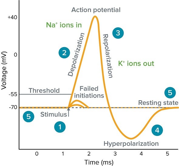
Threshold Potential and Depolarization
- When a stimulus is strong enough, it causes a localized change in membrane potential, depolarizing the membrane to a threshold value, typically around -55 mV.
- Opening of Voltage-Gated Sodium Channels: Once the threshold is reached, voltage-gated sodium channels open, allowing Na⁺ ions to rush into the cell due to the electrochemical gradient.
- The rapid influx of Na⁺ causes the membrane potential to rise quickly from -70 mV to around +30 mV. This shift in voltage is the depolarization phase of the action potential.
Peak of Action Potential
- At the peak of the action potential (around +30 mV), the membrane potential is now positive inside relative to the outside.
- Inactivation of Sodium Channels: Voltage-gated sodium channels close quickly after opening, preventing further Na⁺ influx.
- Opening of Voltage-Gated Potassium Channels: Simultaneously, voltage-gated potassium channels open, allowing K⁺ ions to move out of the neuron.
Repolarization
- As K⁺ ions exit the cell, the membrane potential begins to return to its negative resting state, a process known as repolarization.
- This outflow of positive K⁺ ions restores the negative charge inside the neuron.
- Repolarization brings the membrane potential back toward -70 mV, reversing the earlier depolarization.
Hyperpolarization
- The voltage-gated potassium channels remain open a little longer than necessary, causing an overshoot in K⁺ efflux. As a result, the membrane potential becomes more negative than the resting potential, around -80 to -90 mV. This is known as hyperpolarization.
- During this period, the neuron is in a refractory period:
- Absolute Refractory Period: The neuron cannot initiate another action potential, regardless of stimulus strength, because sodium channels are inactivated.
- Relative Refractory Period: The neuron can fire another action potential, but it requires a much stronger stimulus due to the hyperpolarized state.
Return to Resting Membrane Potential
- After hyperpolarization, the voltage-gated potassium channels close, and the sodium-potassium pump (Na⁺/K⁺ ATPase) helps restore the resting membrane potential of -70 mV.
- The resting potential is maintained until the neuron is ready to fire another action potential.
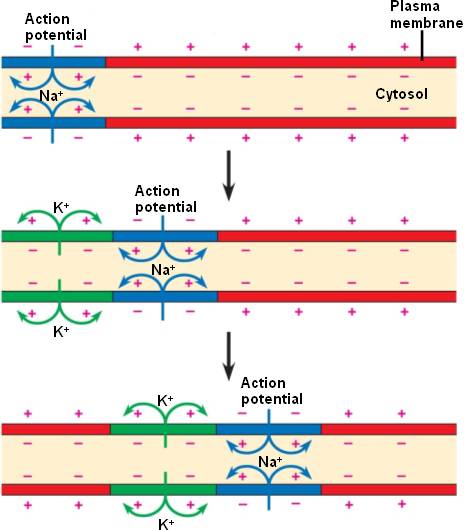
Propagation of Action Potential (Conduction)
Once an action potential is generated at the axon hillock (the region where the axon joins the cell body), it travels along the axon to the axon terminals. This movement of the action potential is called propagation or conduction.
a. Continuous Conduction in Unmyelinated Axons
- In unmyelinated neurons, the action potential travels continuously along the axon, with each successive region of the membrane depolarizing to threshold and generating its own action potential.
- Local Current: The influx of Na⁺ in one region causes adjacent regions of the membrane to reach threshold, opening voltage-gated sodium channels in the next section of the axon. This creates a wave of depolarization that moves down the axon.
- The action potential does not move backward due to the refractory period, ensuring unidirectional movement toward the axon terminal.
b. Saltatory Conduction in Myelinated Axons
- In myelinated neurons, the axon is covered by a fatty substance called myelin, produced by Schwann cells (in the peripheral nervous system) or oligodendrocytes (in the central nervous system). Myelin acts as an insulator, preventing ion exchange across the membrane.
- Nodes of Ranvier: These are gaps in the myelin sheath where voltage-gated sodium and potassium channels are concentrated. The action potential jumps from one node to the next in a process called saltatory conduction.
- Saltatory conduction is much faster than continuous conduction, as the action potential effectively skips over the myelinated sections, only being regenerated at the nodes of Ranvier.
Synaptic Transmission
- Once the action potential reaches the axon terminals, it triggers the release of neurotransmitters into the synaptic cleft.
- Voltage-Gated Calcium Channels: The arrival of the action potential opens voltage-gated calcium channels at the axon terminal, allowing Ca²⁺ ions to enter the neuron.
- Exocytosis of Neurotransmitters: The influx of calcium causes synaptic vesicles to fuse with the membrane and release neurotransmitters into the synapse.
- These neurotransmitters bind to receptors on the postsynaptic neuron, muscle, or gland, which can trigger an action potential or other response in the receiving cell.
All-or-None Principle
- The action potential follows the all-or-none principle, meaning that once the threshold is reached, an action potential will be generated, and it will always have the same magnitude and duration. A stronger stimulus does not produce a stronger action potential but may increase the frequency of action potentials.
Speed of Action Potential Conduction
Several factors affect the speed at which an action potential travels along an axon:
- Axon Diameter: Larger diameter axons have lower internal resistance to the flow of ions, allowing action potentials to propagate faster.
- Myelination: Myelinated axons conduct action potentials much faster than unmyelinated ones due to saltatory conduction.
- Unmyelinated Axons: Conduct at around 1 m/s.
- Myelinated Axons: Can conduct at speeds of up to 120 m/s.
Conclusion
The generation and conduction of action potentials are essential for neuronal communication. This process begins with the depolarization of the neuron’s membrane, followed by the rapid influx of sodium ions, the outflow of potassium ions, and the restoration of the resting potential. The action potential is propagated along the axon to the synapse, where neurotransmitters are released to transmit the signal to the next cell. Myelination and axon diameter play critical roles in the speed of conduction, with myelinated axons exhibiting faster transmission due to saltatory conduction. This mechanism underlies everything from reflex actions to complex thought processes.
Synaptic Transmission
A synapse is a gap that is present between two neurones. Action potentials are propagated across the synapse by synaptic transmission, also known as neurotransmission. The neurone that sends the signal is the presynaptic neurone, whilst the postsynaptic neurone receives the signal.
Neurotransmission starts with the release of a readily available neurotransmitter from the presynaptic neurone, followed by its diffusion and binding to the postsynaptic receptors. Then the postsynaptic cell responds according to the neurotransmitter. Following this, the neurotransmitter is removed or deactivated, allowing the entire cycle to occur again.
In this article, we shall look at the stages of synaptic transmission and clinical conditions that arise in its pathology.

Fig 1 – Diagram showing the basic model of neurotransmission. (A) Presynaptic neuron. (B) Postsynaptic neuron. (1) Mitochondria. (2) Synaptic vesicles containing neurotransmitters. (3) Autoreceptor. (4) Synaptic cleft. (5) Neurotransmitter receptor. (6) Calcium channel. (7) Fused vesicle releasing neurotransmitter. (8) Neurotransmitter reuptake pump.
Synthesis and Storage of NeurotransmittersThis is the first step of synaptic transmission. Some neurotransmitters (eg acetylcholine, ACh) are synthesised in the axon, while others (eg neuropeptides) are made in the cell body.
- Acetylcholine – This is synthesised within the synaptic terminal of the axon. Its precursors (choline, acetate) are taken into the cell by membrane channels or created as byproducts of other processes. Enzymes (such as choline acetyltransferase) convert precursors into the neurotransmitter.
- Endogenous opioids (eg. enkephalins) – These are an example of neuropeptides. These large neurotransmitters are produced within the cell body via transcription in the nucleus and translation in the endoplasmic reticulum. Synthesised precursors are then packaged into secretory granules and sent to the axonal terminal. Importantly, proteases present in the granules cleave the precursors into their mature neuropeptide form during this journey.
Once synthesised, neurotransmitters are stored in vesicles within the synaptic terminal until an action potential arrives, causing their release. Neurotransmitters such as acetylcholine are stored within the small synaptic vesicles, whereas neuropeptides reside within large dense-core vesicles.
Neurotransmitter ReleaseAction potentials depolarising the synaptic terminal lead to the opening of voltage-gated calcium channels. This allows an influx of calcium in the terminal and fusion of the synaptic vesicles with the cell membrane (exocytosis). Consequently, the neurotransmitter is released into the synaptic cleft.
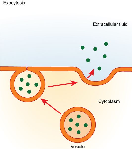
Fig 2 – Diagram showing exocytosis, the process by which neurotransmitters are released into the synaptic cleft.
Postsynaptic ReceptorsThe neurotransmitter in the synaptic cleft diffuses across the gap to the post-synaptic membrane. Here, they can bind to two types of post-synaptic receptors.
| Name | Inotropic receptors | Metabotropic receptors |
| Type | Ligand-gated ion channels | G protein-coupled receptors |
| Response | Channel allows ion flux to change the cellular voltage | Receptor acts through secondary messengers to cause cellular effects |
| Speed of response | Rapid | Slow |
| Length of response | Short-acting | Prolonged response |
This can cause either depolarisation to promote or hyperpolarisation to inhibit the action potential generation in the post-synaptic neurone.
Inactivation/Removal of NeurotransmittersOnce the post-synaptic membrane has responded to the neurotransmitter in the synaptic cleft; it is either inactivated or removed. This can be done in several ways:
- Re-uptake – serotonin is taken back into the pre-synaptic neurone by the transporter proteins in the neuronal membrane. These neurotransmitters are subsequently either recycled by re-packaging into vesicles or broken down by enzymes.
- Breakdown – acetylcholine is broken down by acetylcholinesterase present in the synaptic cleft, inactivating the neurotransmitter.
- Diffusion – into surrounding areas
Neuromuscular Transmission
The intersection of a muscle fibre and a nerve fibre is known as a neuromuscular junction. Neuromuscular transmission is the process by which information is transmitted from a motor nerve ending to a muscle fibre via a neuromuscular junction.
Neuromuscular Transmission
The definition of neuromuscular transmission is the information flow from the motor nerve ending to the muscle fibre via the neuromuscular junction. It is the process by which the motor nerve impulses start the contraction of the muscles.
The neuromuscular junction experiences a number of events throughout this process. Events include:
1. Acetylcholine release
2. Acetylcholine’s action
3. Increasing endplate potential
4. Potential for a miniature endplate development
5. Acetylcholine is destroyed
Neuromuscular Transmission – Steps
The five important steps in the process of neuromuscular transmission are as follows:
1. Acetylcholine release – When an action potential reaches an axon terminal, it opens voltage-gated calcium channels in the axon terminal’s membrane. The axon terminal receives calcium ions from the extracellular fluid. These force the synaptic vesicles to move and fuse with the presynaptic membrane and result in the bursting of the vesicles. Acetylcholine is now released from the ruptured vesicles. Acetylcholine diffuses through the presynaptic membrane and enters the synaptic cleft through the process of exocytosis.
2. Acetylcholine’s action – Acetylcholine molecules enter the synaptic cleft, where they interact with nicotinic receptors in the postsynaptic membrane to form the acetylcholine-receptor complex. By opening the ligand-gated sodium channels, it increases the postsynaptic membrane’s permeability for sodium. Now, sodium ions from ECF pass through these channels and enter the neuromuscular junction. And there, sodium ions change the electrical potential and create the endplate potential.
3. Increasing end plate potential – End plate potential is the shift in resting membrane potential that occurs at the neuromuscular junction as a result of an impulse. End plate potential is the result of a slight depolarization that happens when sodium ions enter the inside.
4. Potential for a miniature endplate development – Miniature endplate potential, which develops when a small amount of acetylcholine is released from the axon terminal, is a weak endplate potential in the neuromuscular junction. The muscle cannot develop an action potential as a result. The miniature endplate potentials are eventually added together to produce endplate potential, which results in an action potential in the muscle.
5. Acetylcholine is destroyed – Acetylcholinesterase enzyme quickly destroys the acetylcholine released into the synaptic cleft. The rapid breakdown of acetylcholine has some functional significance. It stops the muscle fibre from being excited repeatedly and enables the muscle to relax.

Neuromuscular Junction Flowchart
Nerve impulse or action potential
↓
Opening of voltage-gated calcium channels
↓
Influx of calcium ions inside the cell increases
↓
Opening/rupture of vesicle and release of acetylcholine
↓
Acetylcholine comes to the synaptic cleft
↓
Acetylcholine binds with nicotinic receptors to form the acetylcholine-receptor complex
↓
Opening of ligand-gated sodium channels
↓
Influx of sodium ions inside the cell increases
↓
Development of endplate potential
↓
Generate muscle action potential
↓
Muscle contraction takes place